
Medical Information
Transposition of the Great Vessels
1) "Red blood" pumped continually through the left side of the heart to the lungs and back, without entering the rest of the body.
2) "Blue blood" pumped continually through the right side of the heart to the body and back, without entering the lungs.
Babies born with transposition are cyanotic (bluish color of skin, lips, and nail beds) shortly after birth because of the low oxygen in their blood. Two normal connections in the newborn heart and blood vessels help some "red blood" and "blue blood" to mix, keeping babies alive. One connection, the foramen ovale, is an opening between the two atria (upper chambers). In some patients, if the foramen ovale is not open enough for blood to mix, a balloon can be passed through this opening, making it larger. In addition, medicine can be given to keep the second connection (called a patent ductus arteriosus) open. This second connection is a blood vessel between the aorta and pulmonary artery, which usually closes after the first few days of life.
Ultimately, surgery is required in order for enough red blood to circulate through the body. The type of surgery performed depends upon a number of factors including the age of the child, the child's general state of health and the exact nature of the heart defect.
Children with transposition require penicillin (also called SBE prophylaxis) prior to any dental work or surgery on the mouth, bowel, or bladder. This helps prevent the uncommon, but possible, occurrence of bacterial infection affecting the heart near the defect or valves.
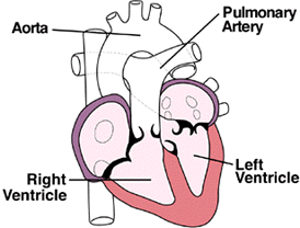
NORMAL HEART![]()
TRANSPOSITION
OF THE GREAT VESSELS
In the normal heart, their are two "great vessels": the aorta which carries blood from the left ventricle to the body and the pulmonary artery which carries blood from the right ventricle to the lungs. In transposition, these two great vessels are switched (transposed) with the aorta arising from the right ventricle, and pulmonary artery arising from the left ventricle. This results in two separate circulations:
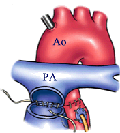
Balloon Atrial Septostomy
What is a Balloon Atrial Septostomy?
A catheter is a special thin tube passed into the blood vessels through a small needle-stick in the groin or forearm, and guided into the heart. Through this catheter, a special device that resembles a balloon is passed into the heart. The wall between the right and left atrium is punctured and the catheter device pushed through the small hole thus created. The balloon is then inflated, and the catheter is pulled back through the small hole, tearing it and making it larger. This procedure is called a Balloon Atrial Septostomy and was first devised by Dr.Rashkind. The aim is to create a large enough opening between the two atria, so that blood can freely mix across it, and improve oxygen supply.
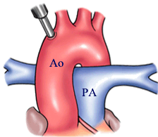
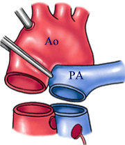
How is an Arterial Switch Operation (ASO) done?
Surgery is carried out through an opening in the middle of the chest. The heart will have to be stopped temporarily during the operation. So the surgeon will first hook up the patient to the heart-lung machine. The aorta and pulmonary artery are disconnected from their abnormal attachments. Their positions are then "SWITCHED". The aorta is stitched back to the left ventricle and the pulmonary artery to the right ventricle. A VSD is closed, if present. The coronary arteries are also freed, and connected back to the aorta using very delicate hair-thin sutures. When you consider that the size of these coronary arteries in a new-born is hardly a millimeter, you can imagine the technical skill and expertise that the surgeon must possess to carry out this connection without mishap.
What is the long term outcome after Arterial Switch Operation?
When an ASO is performed well, the mortality risk is minimal, and long term survival is excellent. Essentially all patients are active without any limitations. Although the longest follow-up is barely 20 years, it is predicted that about 90% of operated patients will survive to this period.
Late complications are uncommon. Rare problems are obstruction at the site of connection of the pulmonary artery to the right ventricle, producing Pulmonary Stenosis, and sometimes leak of the new aortic valve - Aortic Regurgitation. A second operation has hardly ever been necessary for these complications. (However, Ryan underwent a second surgery at 1 year old.)
 |
 |
 |
Sometimes, this pulmonary valve is extremely narrow, and blocks the smooth flow of blood into the lungs. This condition is called Pulmonary Stenosis - or PS, in short. Occassionally, there may be a narrowing of the pulmonary artery or its branches above the pulmonary valve. This is called "supra-valvular" pulmonary stenosis.
What happens in PS ?
What needs to be done for PS ?
What are the options for treating PS ?
What operations are performed for PS ?
Additional sites of block are sought. If there is an obstruction below the valve due to abnormal muscle bundles, these are divided and removed. If the pulmonary artery or its branches are narrow, they are widened by using a patch of pericardium to increase their size - similar to the repair used in Tetralogy of Fallot.
Sometimes the valve may be extremely narrow and this alone is not enough. In such cases, a Trans-Annular Patch repair may be indicated. In this repair, a cut is made across the narrow area, and the opened up portion is roofed using a patch of pericardium or synthetic fabric.
Other defects that occur in addition to PS need correction at the same operation. The commonest of these are ASD and PDA.
What are the risks of surgery ?
What about the long term effects ?
The word "pulmonary" denotes "to do with the lungs". The pulmonary valve is located between the right ventricle and the pulmonary artery. It regulates blood flow into the lungs, and prevents blood from leaking back into the right ventricle.
In PS, blood flow to the lungs is reduced. So the amount of blood getting oxygenated is less than normal. In patients who have very slight narrowing, there is not much effect. But when the block becomes severe, symptoms begin. Usually children with pulmonary stenosis have a limited tolerance to exercise. They become tired easily at play, and have to rest. Rarely, giddiness and fainting may occur. Some children complain of palpitations - which is an uncomfortable awareness of their own heart-beat. In very severe PS, there may be mild cyanosis - a bluish discoloration of the body. The cyanosis is caused by veno-arterial mixing across the wall between the right and left atrium. The diagnosis usually is obvious on medical examination. Confirmation may be obtained by tests like echocardiography, or sometimes by cardiac catheterization.
In PS, the smooth flow of blood into the lungs is obstructed. This needs to be relieved. In the early stages of the disease, not many effects are seen. But if left untreated for many years, it may cause problems. The right ventricle, which pumps blood into the lungs, is now forced to work harder against the blocked pulmonary valve. To do this, the right ventricle wall becomes thicker by addition of more muscle - a condition called "hypertrophy". But even the stronger right ventricle cannot keep up this hard work forever. Finally, it "fails" to pump blood effectively, producing sudden worsening of symptoms. At this stage, urgent treatment becomes necessary, and carries a higher risk.
So although PS is usually not an emergency, it is advisable to seek treatment early rather than delaying the process till complications set in.
For many years surgery has been the only effective treatment for PS. But with the arrival on the scene of catheter-based therapy, it has today virtually taken-over the treatment of PS. Trans-Catheter Balloon dilatation is effective in most cases, and has lesser morbidity than surgery.
The common procedure is called Open Pulmonary Valvotomy (OPV). This is an "open-heart" operation, and is done after the patient is hooked up to a heart-lung machine. The pulmonary artery is opened, and the narrow pulmonary valve examined. Usually, the leaflets of the pulmonary valve are stuck to the wall of the pulmonary artery - "tethering" - and to one another along well-defined lines - "cuspal fusion". The valve is opened-up by dividing its leaflets along the lines of attachment using a scalpel.
Surgery for PS is relatively safe. The complication rate is very low, and includes those common for any open-heart operation. This includes bleeding needing blood transfusions and rhythm disturbances - arrhythmia. Sometimes the relief of block may be incomplete due to technical problems. Most types of PS however can be effectively relieved by operation. Also, any additional defects can be treated at the same time.
Most patients operated for pulmonary stenosis go on to lead normal lives. The risk of late complications is very low. In most children, as growth occurs, the pulmonary valve also becomes larger, and this itself reduces the stenosis to some extent. The potential problems are rhythm disturbances and "late" pulmonary valve leak due to injury at the time of operation.
What is Cardiac Catheterization and how is it performed?
During cardiac catheterization, a thin tube called a catheter is fed through a blood vessel to a part of the body that needs to be assessed. The catheter is inserted through a very small cut made by the physician (in the groin, arm or wrist), then guided up through the blood vessel to the heart. The physician tracks the course of the catheter by watching it on a fluoroscope, which displays the blood vessels on a viewing screen. A variety of measurements are performed when the catheter is in place, and then the catheter is removed. After some recovery time, most patients are free to go home after about six hours. Results should be available within a matter of hours. Back to updates
What is Angioplasty and how is it performed?
Angioplasty may be done by attaching a small balloon to the catheter. Once the catheter has been guided to the proper location in an artery, the balloon is inflated. The pressure from the inflated balloon presses fat and calcium deposits (plaque) against the wall of the artery to improve blood flow.
What is Stenting?
First performed in the mid-1980s, and first approved by the FDA in the mid 1990s, stenting is a catheter-based procedure in which a stent (a small, expandable wire mesh tube) is inserted into a diseased artery to hold it open. Currently, stenting is performed most often in conjunction with other catheter-based procedures, such as balloon angioplasty or atherectomy. The other procedures are used to partially reduce the narrowing caused by atherosclerosis, and the stent typically allows for an excellent final result to be obtained with little to no narrowing remaining within the coronary arteries. By doing a stent insertion along with these other procedures, the risk of the artery re-closing (restenosis) is reduced, and the risk of abrupt vessel closures during or within 24 hours of the procedure is nearly eliminated.
Within one month, the stent becomes covered with a thin layer of the artery’s inner lining cells. It will not be affected by any mechanical equipment or detected by a metal detector. The success of a stenting procedure can be threatened by risk factors such as smoking or high cholesterol levels, which unchecked could lead to new blockages in the coronary arteries. Therefore, people receiving stents are strongly encouraged to learn and practice healthy lifestyle behaviors for good heart health.
Sinus Node Dysfunction
What is Sinus Node Dysfunction?
Symptoms of Sinus Node Dysfunction
When symptoms do occur, they may include:
Causes of Sinus Node Dysfunction
Types of Sinus Node Dysfunction
What makes the Sinus Node misfire?
Sick sinus syndrome — also known as SINUS NODE DYSFUNCTION — is the name for a group of heart rhythm problems (arrhythmias) in which the sinus node — the heart's natural pacemaker — doesn't work properly. The sinus node is an area of specialized cells in the upper right chamber of the heart that controls the rhythm of your heart. Normally, the sinus node produces a steady pace of regular electrical impulses. In sick sinus syndrome, these signals are abnormally paced. A person with sick sinus syndrome may have heart rhythms that are too fast, too slow, punctuated by long pauses — or an alternating combination of all of these rhythm problems. Sick sinus syndrome is relatively uncommon, but the risk of developing sick sinus syndrome increases with age. Many people with sick sinus syndrome eventually need a pacemaker to keep the heart in a regular rhythm.
Most people with sick sinus syndrome initially have few or no symptoms. In some cases, symptoms may come and go. Many of these signs and symptoms are caused by reduced blood flow to the brain when the heart beats too fast or too slowly.
•Slower than normal pulse (bradycardia)
•Fatigue
•Dizziness or lightheadedness
•Fainting or near fainting
•Shortness of breath
•Chest pains
•A sensation of rapid, fluttering heartbeats (palpitations)
Your heart is made up of four chambers — two upper chambers (atria) and two lower chambers (ventricles). The rhythm of your heart is normally controlled by the sinoatrial (SA) node — or sinus node — an area of specialized cells located in the right atrium. This natural pacemaker produces the electrical impulses that trigger each heartbeat. From the sinus node, electrical impulses travel across the atria to the ventricles, causing them to contract and pump blood out to your lungs and body. If you have sick sinus syndrome, your sinus node isn't functioning properly, so your heart rate may be too slow (bradycardia) or too fast (tachycardia) or irregular.
Types of sick sinus syndrome and their causes include:
•Sinoatrial block. Electrical signals move too slowly through the sinus node, causing an abnormally slow heart rate.
•Sinus arrest. The sinus node activity pauses.
•Bradycardia-tachycardia syndrome. The heart rate alternates between abnormally fast and slow rhythms, usually with a long pause (asystole) between heartbeats.
Diseases and conditions that cause scarring or damage to your heart's electrical system can be the reason. Scar tissue from a previous heart surgery also may be the cause, particularly in children. Sick sinus syndrome may also be set off by medications, such as calcium channel blockers or beta blockers used to treat high blood pressure, heart disease or other conditions. However, in many cases, the sinus node doesn't work properly because of age-related wear and tear to the heart muscle.